
Humans possess the ability to walk in a variety of situations – from navigating through crowds to traversing tightropes. However, due to limitations in brain imaging technology, the inner workings of the brain during adaptation of walking patterns have largely remained a mystery.
“You walk in different ways all the time. You deal with terrain, you deal with speed, you deal with dodging other people…but we don’t know how your brain manages it,” said Dr. Daniel Ferris, Robert W. Adenbaum Professor & senior associate chair of the J. Crayton Pruitt Family Department of Biomedical Engineering at the University of Florida.
The National Institutes of Health has recently awarded Ferris $2 million to investigate how the human brain controls locomotor adaptation under varying conditions. Information gained from this study could aid in developing new methods of diagnosis and treatment for gait disabilities.
Conventional methods of brain imaging, such as MRI, require subjects to be stationary, as signals become noisy and difficult to read in motion. These methods also typically record too slowly for brain activity to be identified within a single step.
To overcome the drawbacks of previous technology, Ferris and his lab modified electroencephalography or EEG, sensors to better cancel noise from head motion.
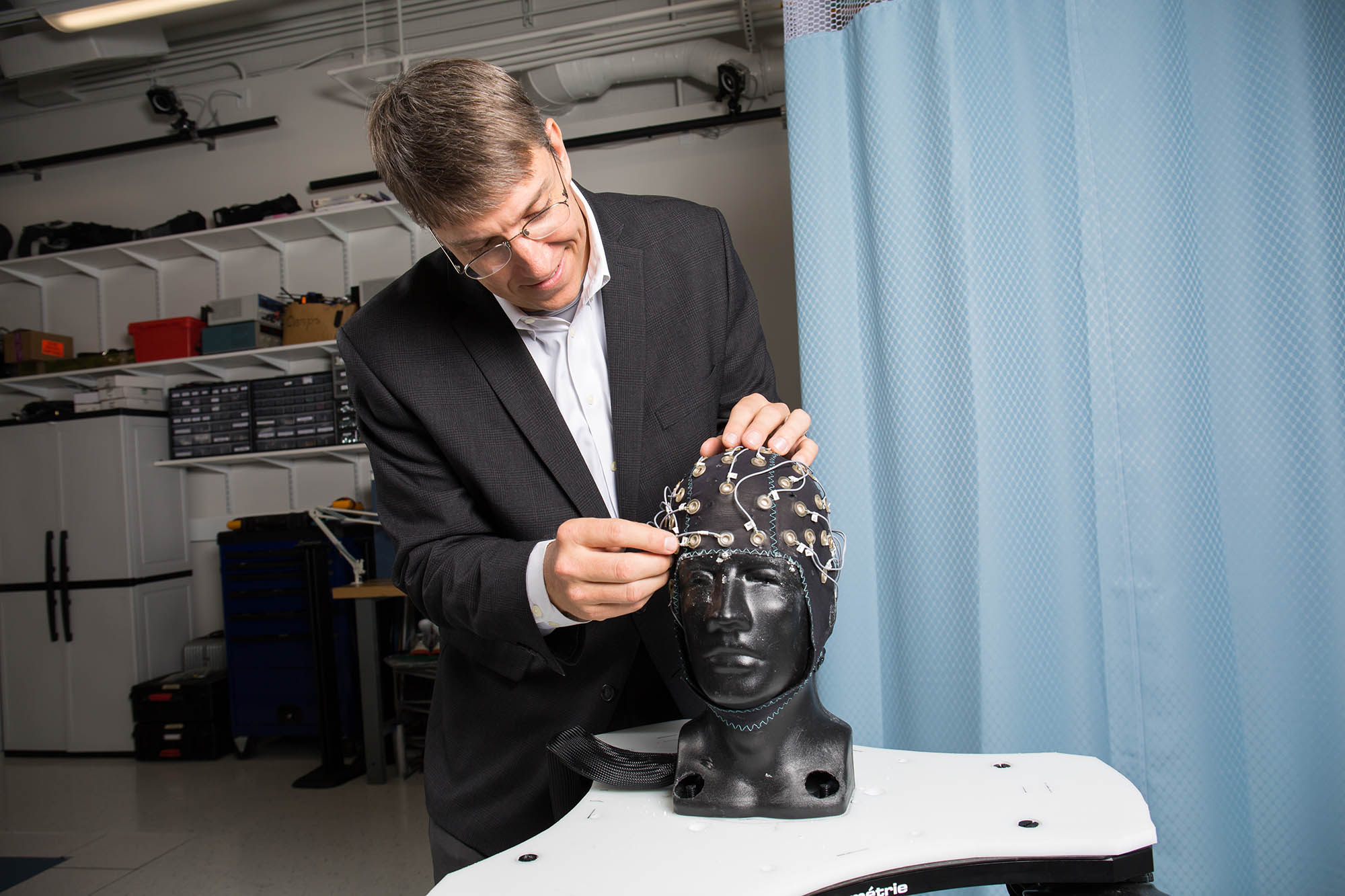 “We use high-density (EEG), meaning we use many electrodes in a small area… we put 256 electrodes on your head and we record many different places,” Ferris said.
To accurately detect rapid changes in brain activity of subjects in motion, this system utilizes new noise-canceling electrodes and signal processing techniques, Ferris said.
While walking on treadmills, subjects will wear an EEG headset so their brain activity is recorded as they adapt their gait to increasing speeds. An additional condition will cause patients to limp as they walk on a split-treadmill where the two treads move at different rates.
Another experiment will involve the strengthening of one ankle with a robotic exoskeleton developed in Ferris’s lab. Existing exoskeletons have been designed for disabled patients but it is unknown how the brain adapts to them, Ferris said.
“It makes you stronger. Every time you push off (with your foot) when you’re walking, you get not only your normal torque but you get extra torque,” Ferris said.
By using virtual reality, the effects of disturbing visual input during walking will also be investigated.
“(Researchers) have found that if you blindfold stroke patients during rehab, it helps them improve their balance,” Ferris said. “The idea is if you take away your visual feedback, you have to rely on your proprioception, your sense of where your body is.”
Virtual reality goggles placed on subjects will display a representation of their environment, but about 10% of the time the display will be rotated, causing subjects to trust their vision less.
Upcoming studies will focus on healthy patients to elucidate the details of normal brain function. In the future, however, similar experiments could reveal how conditions such as aging, stroke, or spinal cord injury affect locomotor adaptation in patients.
By Jonathan Griffin, BME Ph.D. student