
Dr. Edward Phelps, assistant professor in the J. Crayton Pruitt Family Department of Biomedical Engineering, is a co-author of an article in Scientific Reports, an open-access journal from the Nature Publishing Group.
The paper titled, “Advances in pancreatic islet monolayer culture on glass surfaces enable super-resolution microscopy and insights into beta cell ciliogenesis and proliferation” describes a novel method to advance high-resolution imaging of live or fixed beta cells as well as a protocol to induce beta cell proliferation. The aim of this paper is to investigate a new method of culturing islet cells that improves imaging capabilities, thus enabling us to better study subcellular-level factors relevant to type 1 and type 2 diabetes.Understanding the cell biology in health and disease of insulin-producing beta cells in pancreatic islets is central to developing novel therapies for the treatment of both type 1 and type 2 diabetes. The field of pancreatic islet cell biology has long been hampered by insufficient techniques to culture primary insulin-producing beta cells on the surface of glass coverslips for manipulation and high-resolution imaging. Phelps and co-first author, Chiara Cianciaruso, a postdoctoral scientist at the Baekkeskov Laboratory at the Swiss Federal Institute of Technology in Lausanne, Switzerland, made the fundamental discovery that primary pancreatic beta cells are able to grow on glass surfaces when treated to culture conditions and growth media developed for neurons. Using this method, the authors were able to establish the first convincing examples of super-resolution light microscopy of primary human beta cells on glass. Phelps and colleagues demonstrate the applicability of the new islet cell culture method towards studying a novel mechanism to promote the proliferation of primary beta cells. Phelps expects that the new method for culturing monolayers of primary islet cells will become important for a variety of studies of islet cell biology. The method will also be of interest to other fields studying difficult-to-culture primary cells.
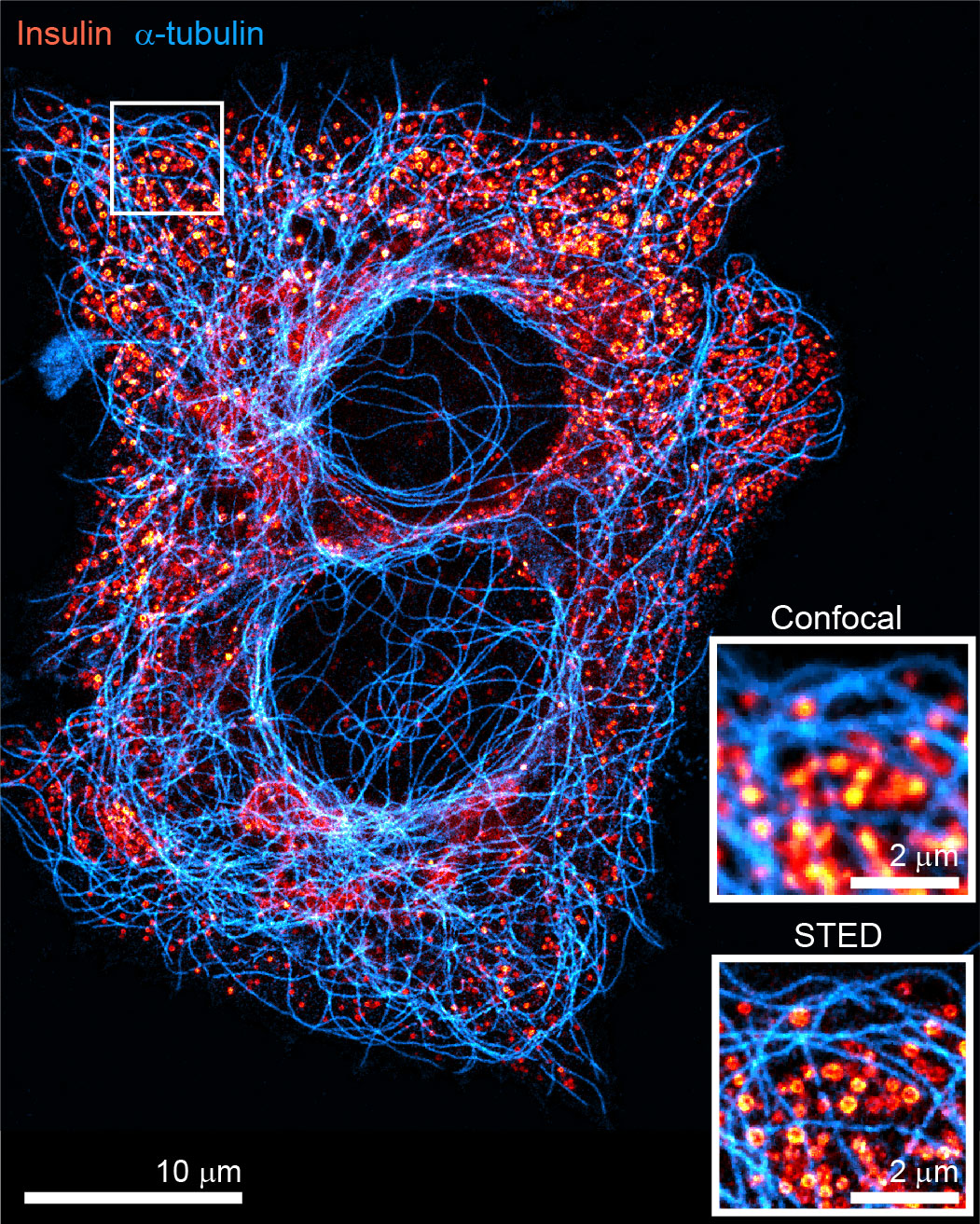 Image caption:
Using culture techniques typically reserved for neurons, Phelps and colleagues grew primary human beta cells on the surface of glass coverslips which enabled them to image the beta cells by stimulated emission depletion (STED) super-resolution light microscopy. Features such as the depicted microtubule cytoskeleton and insulin secretory granules were able to be resolved in much greater detail than is possible by traditional confocal microscopy.