
Dr. Wesley Bolch and researchers were awarded an NIH National Institute of Allergy and Infectious Diseases program project grant for $10.7 million to conduct multiscale evaluations and to investigate mitigation of toxicities following internal radionuclide contamination.
Wesley Bolch, PhD, (PI) Distinguished Progressor and UF Term Professor will lead the efforts for the University of Florida. The UF Project 1 is one of four efforts in this P01 grant. Other projects include those at The Georgia Institute of Technology (Project 2), Northwestern University (Project 3), and the University of California-Berkeley (Project 4). Gayle E. Woloschak, PhD, Professor of Radiation Oncology at Northwestern University, will lead serve as overall Project Director of the NIAID Program.
Bolch along with UF co-investigators Ruogu Fang, PhD, Associate Professor in the Department of Biomedical Engineering, and John Aris, PhD, Associate Professor in the Department of Anatomy and Cell Biology will focus on developing tools and approaches for the rapid assessment of levels of internal radionuclide contamination in members of the general public following the accidental or intentional release of radioactive aerosols.
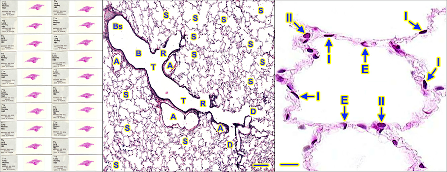
Image details: Construction of a microscale histology-based anatomic model of the lungs: Lung H&E: (Left) 20 of 77 slides from tissue block 71A. (Middle) A, pulmonary artery; B, bronchiole; Bs, bronchus (segmental); D, alveolar duct; R, respiratory bronchiole; S, alveolar sac; T, terminal bronchiole; bar, 200 µm. (Right) E, endothelial cells (lining capillary of the alveolar wall); I, type I alveolar cell; II, type II alveolar cell; bar, 20 µm.
Radiological terrorism events can involve two broad classes of devices. The first, and perhaps the most likely, would be a Radiological Dispersion Device (RDD, commonly referred to as a “dirty bomb”). An RDD is composed of a traditional explosive incorporating either encapsulated or free forms of a radionuclide or possibly a radionuclide mixture. The main impact on those civilians near the device will include bodily injury due to the initial explosive shock wave, ejected shrapnel, and induced skin burns.
Depending upon the location of the device, its explosive yield, and the chemical form of the radionuclide, the main subsequent impact to the majority of victims will be inhalation of air-dispersed radionuclide aerosols. Within minutes to hours, radionuclide intakes may additionally include inhalation of resuspended particles.
The second form of a radiological device, perhaps less likely but far more impactful, would be an Improvised Nuclear Device (IND). Depending upon the location and height of the detonation (hypocenter), the local terrain, and the distribution of the populace relative to the hypocenter, the first impact on victims will be the immediate irradiation of tissues and organs.
Work proposed in this research will provide in-field radiological response tools for organ-level dose assessment given the unique body morphometry of the adult victim. It will extend this ability to other critical members of the general public, including children, adolescents, and pregnant females.
This project aims to develop field-deployable software which, to together with external detector measurements, will permit triage-level reporting of organ dose to individuals internally contaminated with radionuclides following Radiological Dispersion Device (RDD), Improvised Nuclear Device (IND), or Nuclear Reactor Accident (NRA) release.
The University of Florida will be responsible for the following contributions to the grant aims:
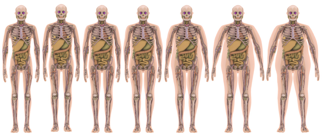
- Develop whole-body scalable tetrahedral mesh computational phantoms of adults, children, and pregnant females to include inter/intra-organ blood vessels for Monte Carlo radiation transport simulations.
- Develop a comprehensive set of detector response functions for hand-held detectors across the full complement of adult, pediatric, and pregnant female computational phantoms developed in SA1.
- Design and construct a single-screen software code to rapidly assess organ doses to RDD/IND/NRA victims following radionuclide intake and validate dose estimates with physical measurements.
- Construct 3D mesh-based histology models of the lung airways, liver, spleen, and bone marrow. Apply x-ray fluorescence microscopy (XFM) imaging data from Northwestern University and UC-Berkeley to model non-uniform radionuclides distributions at the cellular level.